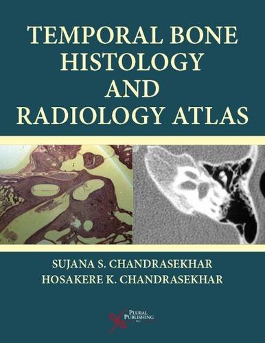
Product Title: Temporal Bone Histology and Radiology Atlas (Original PDF from Publisher)
Format:
Publisher PDF, File Size = 72.90 MB
Overview (Details, Topics and Speakers):
by Sujana S. Chandrasekhar (Author), Hosakere K. Chandrasekhar (Author)
Temporal Bone Histology and Radiology Atlas covers temporal bone histology with radiologic correlates. It examines horizontal and vertical histologic sections of the temporal bone and correlates that microanatomy with that which is seen on CT and MR imaging. This enables the reader to ”see” much more when they look at radiographs than they otherwise would. This text is easy to use and can be referred to briefly and frequently in the course of otolaryngology or radiology practice, and can be digested comfortably for MOC and board preparation.
Key Topics:
- Important anatomical relationships
- Microscopic guidance to evaluate radiographic images
- Special preparation techniques for electron microscopy and DNA extraction
- Routine and special histology techniques
The images in the book are available on a companion site allowing the reader to zoom in for more detail.
Temporal Bone Histology and Radiology Atlas is designed for otolaryngology and radiology residents and fellows, otolaryngologists and radiologists both for clinical practice and in preparation for maintenance of certification and licensure exams, and medical students interested in otolaryngology and radiology.
Product Details
- Hardcover: 230 pages
- Publisher: Plural Publishing, Inc.; 1 edition (January 30, 2018)
- Language: English
- ISBN-10: 9781597567169
- ISBN-13: 978-1597567169
- ASIN: 1597567167
- Product Dimensions: 8.8 x 0.7 x 11.2 inches
Delivery Method
the Temporal Bone Histology and Radiology Atlas (Original PDF from Publisher) course/book will be provided for customer as download link. download link has NO Expiry and can be used anytime.
Contact Us
contact us to our email at [email protected] or fill in the form below:




Reviews
There are no reviews yet.