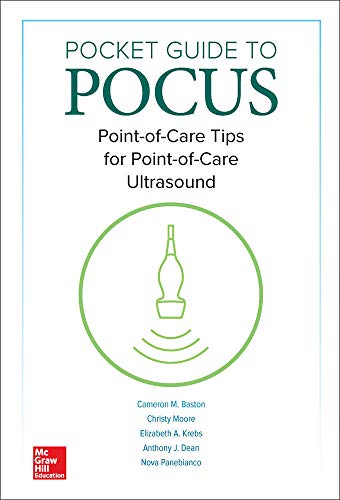
Product Title: Pocket Guide to POCUS: Point-of-Care Tips for Point-of-Care Ultrasound (Videos)
Format:
148 Videos, File Size = 458.00 MB
Overview (Details, Topics and Speakers):
VIDEOS INCLUDED:
- Video 01-01: Cardinal Movements
- Video 02-01: Acoustic Shadowing
- Video 02-02: Posterior Acoustic Enhancement
- Video 02-03: Reverberation Artifact
- Video 02-04: Mirror Artifact
- Video 02-05: Edge Artifact
- Video 02-06: Orientation Check
- Video 02-07: Color Doppler
- Video 03-01: Hand Position Longitudinal
- Video 03-02: Hand Position Transverse
- Video 03-03: Upper Arm Anatomy
- Video 03-04: Forearm Anatomy
- Video 03-05: Accessing the Vessel in Transverse
- Video 03-06: Accessing the Vessel in Longitudinal
- Video 03-07: Needle through the Posterior Wall
- Video 03-08: Lymph Node
- Video 03-09: Superficial Thrombophlebitis
- Video 03-10: Infiltrated Line – Longitudinal
- Video 03-11: Valve
- Video 04-01: Normal RUQ
- Video 04-02: Normal LUQ
- Video 04-03: Normal Subxiphoid
- Video 04-04: Normal Pelvic
- Video 04-05: Abnormal RUQ
- Video 04-06: Abnormal LUQ
- Video 04-07: Abnormal Pelvic – Female
- Video 04-08: Loculated Ascites
- Video 04-09: Abnormal Pelvic – Male
- Video 04-10: Seminal Vesicles
- Video 04-11: LUQ Fluid Filled Stomach
- Video 05-01: Free fluid in right lower quadrant
- Video 05-02: Vessel check with linear transducer
- Video 05-03: Real time needle guidance
- Video 05-04: Vessel check with color Doppler
- Video 05-05: Safe Pocket of Ascites
- Video 05-06: Free fluid compared to fluid in viscera
- Video 05-07: Ascites with liver tip in view
- Video 05-08: Ascites with spleen tip in view
- Video 05-09: Full bladder
- Video 05-10: Loculated ascites
- Video 05-11: Epigastric vessels
- Video 05-12: Shallow ascites with visible stool
- Video 06-01: A lines
- Video 06-02: Lung curtain
- Video 06-03: Lung sliding
- Video 06-04: B lines
- Video 06-05: Consolidation
- Video 06-06: Stratosphere sign
- Video 06-07: Pleural effusion
- Video 06-08: Subpleural consolidations
- Video 06-09: Z lines
- Video 06-10: Shred sign
- Video 07-01: Left lung base
- Video 07-02: Right lung base
- Video 07-03: Rib shadow
- Video 07-04: Depth measurement
- Video 07-05: Small effusion
- Video 07-06: Dense atelectasis
- Video 07-07: Loculated effusion
- Video 07-08: Single loculation
- Video 07-09: Pleural mass
- Video 07-10: Large effusion
- Video 08-01: Parasternal long axis view (PLAX)
- Video 08-02: Parasternal short axis view (PSSA)
- Video 08-03: Apical four chamber view (A4C)
- Video 08-04: Subxiphoid view
- Video 08-05: Transverse view of the Inferior Vena Cava (IVC)
- Video 08-06: Longitudinal view of the Inferior vena cava (IVC)
- Video 08-07: High Ejection fraction PSSA
- Video 08-08: Parasternal long axis view with pericardial effusion
- Video 08-09: Pleural effusion in the parasternal long axis view
- Video 08-10: Low Ejection fraction in the parasternal long axis
- Video 08-11: Tamponade in the parasternal long axis view
- Video 08-12: Right ventricular enlargement in the parasternal long axis view
- Video 08-13: Right ventricular enlargement in the parasternal short axis view
- Video 08-14: Low Ejection fraction in the parasternal short axis
- Video 08-15: Parasternal short axis view with a pericardial effusion
- Video 08-16: Apical four chamber view (A4C) with a low ejection fraction
- Video 08-17: Apical four chamber view (A4C) with a pericardial effusion
- Video 08-18: Apical four chamber view with dilated right ventricle
- Video 08-19: Subxiphoid view with pericardial effusion
- Video 08-20: Transverse view of a dilated inferior vena cava
- Video 08-21: Longitudinal view of a flat inferior vena cava
- Video 09-01: Transverse view of a normal inferior vena cava
- Video 09-02: Longitudinal view of a normal IVC
- Video 09-03: A lines at the apex of normal lungs
- Video 09-04: Longitudinal view of the internal jugular vein
- Video 09-05: Normal parasternal long axis view
- Video 09-06: Normal parasternal short axis
- Video 09-07: Longitudinal view of a flat inferior vena cava
- Video 09-08: Transverse view of a flat inferior vena cava
- Video 09-09: Longitudinal view of a plethoric IVC
- Video 09-10: Transverse view of a plethoric IVC
- Video 09-11: Discrete B lines in a patient with early pulmonary edema
- Video 09-12: Coalescent B lines in a patient with severe pulmonary edema
- Video 09-13: Enlarged left atrium in a parasternal view
- Video 09-14: Decreased left ventricular end diastolic diameter in the parasternal short view
- Video 10-01: Anatomy scan of normal femoral vessels
- Video 10-02: Compression of normal femoral vessels
- Video 10-03: Anatomy scan of normal popliteal vessels
- Video 10-04: Compression of normal popliteal vessels
- Video 10-05: Incompressible femoral vein
- Video 10-06: Incompressible popliteal vein
- Video 10-07: Visible thrombus in multiple vessels
- Video 10-08: Baker’s cyst
- Video 10-09: Lymph node
- Video 10-10: Superficial Thrombophlebitis
- Video 11-01: Transverse view of carotid and internal jugular
- Video 11-02: Transverse view of carotid and internal jugular
- Video 11-03: Longitudinal view of the catheter in the vein
- Video 11-04: Needle accessing the internal jugular vein in the transverse view
- Video 11-05: Needle accessing a vessel in longitudinal axis
- Video 11-06: Wire entering the vessel in longitudinal view
- Video 11-07: Internal jugular vein directly over carotid artery
- Video 11-08: Internal jugular vein net to carotid artery
- Video 11-09: Axillary vein transitioning to subclavian vein
- Video 11-10: Post procedural hematoma and thrombus of the internal jugular vein
- Video 11-11: Collapsing internal jugular vein
- Video 11-12: Overlying sternocleidomastoid muscle body
- Video 11-13: Catheter penetrating the posterior wall of the vessel in the long axis
- Video 12-01: Normal kidney longitudinal image
- Video 12-02: Normal kidney transverse image
- Video 12-03: Normal kidney with color Doppler
- Video 12-04: Full bladder in a female pelvis
- Video 12-05: Foley balloon in empty bladder
- Video 12-06: Full bladder transverse view
- Video 12-07: Mild hydronephrosis
- Video 12-08: Moderate hydronephrosis
- Video 12-09: Severe hydronephrosis
- Video 12-10: Moderate hydronephrosis with color Doppler
- Video 12-11: Atrophic kidney
- Video 12-12: Renal cyst
- Video 12-13: Ureterovesicular junction stone
- Video 12-14: Intrarenal calculi
- Video 13-01: Abscess
- Video 13-02: Cellulitis
- Video 13-03: Abscess with color Doppler
- Video 13-04: Exam glove as probe cover
- Video 13-05: Water bath technique
- Video 13-06: Squish sign in an abscess
- Video 13-07: Inguinal lymph node
- Video 13-08: Foreign body
- Video 14-01: Ocular anatomy
- Video 14-02: Increased optic nerve sheath diameter (ONSD)
- Video 14-03: Vitreous hemorrhage
- Video 14-04: Retinal detachment with macula on
- Video 14-05: Central retinal artery occlusion
- Video 14-06: Papilledema
Product Details
- Publisher:McGraw-Hill Education / Medical; 1st edition (February 15, 2019)
- Language:English
- Paperback:83 pages
- ISBN-10:1260441474
- ISBN-13:978-1260441475
- ISBN-13:9781260441475
- eText ISBN: 9781260441550
- Item Weight:13.3 ounces
- Dimensions:7 x 0.5 x 10 inches
Delivery Method
the Pocket Guide to POCUS: Point-of-Care Tips for Point-of-Care Ultrasound (Videos) course/book will be provided for customer as download link. download link has NO Expiry and can be used anytime.
Contact Us
contact us to our email at [email protected] or fill in the form below:


Reviews
There are no reviews yet.