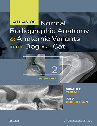
Product Title: Atlas of Normal Radiographic Anatomy and Anatomic Variants in the Dog and Cat, 2nd Edition (Original PDF from Publisher)
Format:
Publisher PDF, File Size = 56.20 MB
Overview (Details, Topics and Speakers):
Equip yourself to make accurate diagnoses and achieve successful treatment outcomes with this highly visual comprehensive atlas. Featuring a substantial number of new high contrast images, Atlas of Normal Radiographic Anatomy and Anatomic Variants in the Dog and Cat, 2nd Edition provides an in-depth look at both normal and non-standard subjects along with demonstrations of proper technique and image interpretations. Expert authors Donald E. Thrall and Ian D. Robertson describe a wider range of “normal” as compared to competing books ― not only showing standard dogs and cats, but also non-standard subjects such as overweight and underweight pets and animals with breed-specific variations. Every body part is put into context with a textual description to help explain why a structure appears as it does in radiographs, and enabling practitioners to appreciate variations of normal that are not included, based on an understanding of basic radiographic principles.
- Radiographic images of normal or standard prototypical animals
- are supplemented by images of non-standard subjects exhibiting breed-specific differences, physiologic variants, or common congenital malformations.
- Images that depict a wider range of “normal” ― such as images that detail the natural growth and aging characteristics of normal pediatric and senior animals ― prevents clinical under- and over-diagnosing.
- In-depth coverage of patient positioning and radiographic exposure guidelines assist clinicians in producing the very best results.
- Unlabeled radiographs along side labeled counterparts clarifies important anatomic structures of clinical interest.
- High-quality digital images provide excellent contrast resolution and better visibility of normal structures to assist clinicians in making accurate diagnoses.
- Brief descriptive text and explanatory legends accompany all images to help put concepts into the proper context.
- An overview of radiographic technique includes the effects of patient positioning, respiration, and exposure factors.
- NEW! Companion website
- features additional radiographic CT scans and more than 100 questions with answers and rationales.
- NEW! Additional CT and 3D images have been added to each chapter to help clinicians better evaluate the detail of bony structures.
- NEW! Breed-specific images of dogs and cats are included throughout the atlas to help clinicians better understand the variances in different breeds.
- NEW! Updated material on oblique view radiography provides a better understanding of an alternative approach to radiography, particularly in fracture cases.
- NEW! 8.5″ x 11″ trim size makes the atlas easy to store.
Product Details
- Publisher:Saunders; 2nd edition (October 13, 2015)
- Language:English
- Hardcover:320 pages
- ISBN-10:032331225X
- ISBN-13:978-0323312257
- ISBN-13:9780323312257
- eText ISBN: 9780323312806
- eText ISBN: 9780323312776
- Item Weight:2.36 pounds
- Dimensions:8.5 x 0.8 x 11 inches
Delivery Method
the Atlas of Normal Radiographic Anatomy and Anatomic Variants in the Dog and Cat, 2nd Edition (Original PDF from Publisher) course/book will be provided for customer as download link. download link has NO Expiry and can be used anytime.
Contact Us
contact us to our email at [email protected] or fill in the form below:

Reviews
There are no reviews yet.