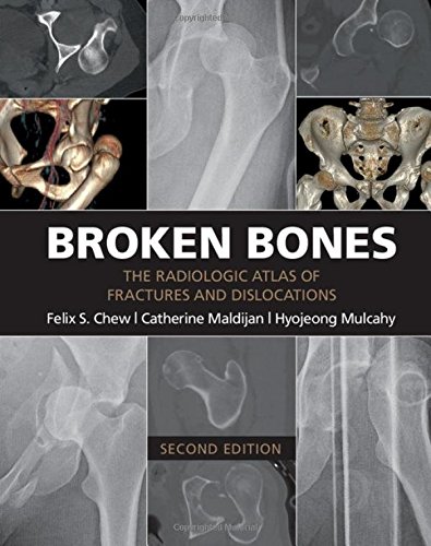
Product Title: Broken Bones: The Radiologic Atlas of Fractures and Dislocations
Format:
Retail PDF,
Overview (Details, Topics and Speakers):
by Felix S. Chew (Author)
Broken Bones contains 434 individual cases and 1,101 radiologic images illustrating the typical and less typical appearances of fractures and dislocations throughout the body. The first chapter describes fractures and dislocations of the fingers, starting with fractures of the phalangeal tufts and progressing through the distal, middle, and proximal phalanges and the DIP and PIP joints. Subsequent chapters cover the metacarpals, the carpal bones, the radius and ulna, the elbow and upper arm, and the shoulder and thoracic cage. The cervical spine and the thoracic and lumbosacral spine are covered in separate chapters, followed by the pelvis, the femur, the knee and lower leg, the ankle, the tarsal bones, and the metatarsals and toes. The final three chapters cover the face, fractures and dislocations in children, and fractures and dislocations caused by bullets and nonmilitary blasts.
Product Details
- Paperback: 392 pages
- Publisher: Cambridge University Press; 2 edition (May 3, 2016)
- Language: English
- ISBN-10: 1107499232
- ISBN-13: 978-1107499232
- Product Dimensions: 8.7 x 0.7 x 11 inches
Delivery Method
the Broken Bones: The Radiologic Atlas of Fractures and Dislocations course/book will be provided for customer as download link. download link has NO Expiry and can be used anytime.
Contact Us
contact us to our email at [email protected] or fill in the form below:


Reviews
There are no reviews yet.