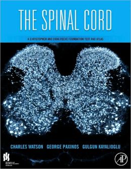
Product Title: The Spinal Cord: A Christopher and Dana Reeve Foundation Text and Atlas (ORIGINAL PDF from Publisher)
Format:
RETAIL PDF, ToC, Index, Full Bookmark,
Overview (Details, Topics and Speakers):
by Charles Watson, George Paxinos (Editor), Gulgun Kayalioglu (Editor), Claire Heise
Many hundreds of thousands suffer spinal cord injuries leading to loss of sensation and motor function in the body below the point of injury. Spinal cord research has made some significant strides towards new treatment methods, and is a focus of many laboratories worldwide. In addition, research on the involvement of the spinal cord in pain and the abilities of nervous tissue in the spine to regenerate has increasingly been on the forefront of biomedical research in the past years. The Spinal Cord, a collaboration with the Christopher and Dana Reeve Foundation, is the first comprehensive book on the anatomy of the mammalian spinal cord. Tens of thousands of articles and dozens of books are published on this subject each year, and a great deal of experimental work has been carried out on the rat spinal cord. Despite this, there is no comprehensive and authoritative atlas of the mammalian spinal cord. Almost all of the fine details of spinal cord anatomy must be searched for in journal articles on particular subjects. This book addresses this need by providing both a comprehensive reference on the mammalian spinal cord and a comparative atlas of both rat and mouse spinal cords in one convenient source. The book provides a descriptive survey of the details of mammalian spinal cord anatomy, focusing on the rat with many illustrations from the leading experts in the field and atlases of the rat and the mouse spinal cord. The rat and mouse spinal cord atlas chapters include photographs of Nissl stained transverse sections from each of the spinal cord segments (obtained from a single unfixed spinal cord), detailed diagrams of each of the spinal cord segments pictured, delineating the laminae of Rexed and all other significant neuronal groupings at each level and photographs of additional sections displaying markers such as acetylcholinesterase (AChE), calbindin, calretinin, choline acetlytransferase, neurofilament protein (SMI 32), enkephalin, calcitonin gene-related peptide (CGRP), and neuronal nuclear protein (NeuN).
The text provides a detailed account of the anatomy of the mammalian spinal cord and surrounding musculoskeletal elements.
The major topics addressed are:
• development of the spinal cord
• the gross anatomy of the spinal cord and its meninges
• spinal nerves, nerve roots, and dorsal root ganglia
• the vertebral column, vertebral joints, and vertebral muscles
• blood supply of the spinal cord
• cytoarchitecture and chemoarchitecture of the spinal gray matter
• musculotopic anatomy of motoneuron groups
• tracts connecting the brain and spinal cord
• spinospinal pathways
• sympathetic and parasympathetic elements in the spinal cord
• neuronal groups and pathways that control micturition
• the anatomy of spinal cord injury in experimental animals
The atlas of the rat and mouse spinal cord has the following features:
• Photographs of Nissl stained transverse sections from each of 34 spinal segments for the rat and mouse.
• Detailed diagrams of each of the 34 spinal segments for rat and mouse, delineating the laminae of Rexed and all other significant neuronal groupings at each level.
• Alongside each of the 34 Nissl stained segments, there are additional sections displaying markers such as acetylcholinesterase, calbindin, calretinin, choline acetlytransferase, neurofilament protein (SMI 32), and neuronal nuclear protein (NeuN).
• All the major motoneuron clusters are identified in relation to the individual muscles or muscle groups they supply.
Product Details
- ISBN-13: 9780123742476
- Publisher: Elsevier Science
- Publication date: 11/26/2008
- Pages: 408
Delivery Method
the The Spinal Cord: A Christopher and Dana Reeve Foundation Text and Atlas (ORIGINAL PDF from Publisher) course/book will be provided for customer as download link. download link has NO Expiry and can be used anytime.
Contact Us
contact us to our email at [email protected] or fill in the form below:


Reviews
There are no reviews yet.