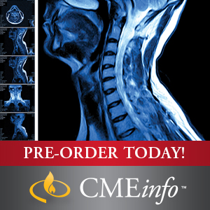
Product Title: UCSF Neuro and Musculoskeletal Imaging 2019 (CME Videos)
Format:
MP4 + PDF, File Size = 7.42 GB
Overview (Details, Topics and Speakers):
UCSF Neuro and Musculoskeletal Imaging
University of California San Francisco Clinical Update (SA-CME)
Stay abreast of the latest developments and trends in neuroradiology and musculoskeletal imaging with this inclusive clinical update.
Stay Current on Rapidly Changing Specialty
UCSF Neuro and Musculoskeletal Imaging is a comprehensive state-of-the-art update on clinically relevant topics in neuroradiology and musculoskeletal imaging. This in-depth CME course includes case-based lectures on topics like skeletal trauma, shoulder instability, brain tumor pitfalls, hydrocephalus, staging cervical nodes, and more. It will help you to better:
- Revamp imaging protocols for brain, head & neck, spine, nerve and musculoskeletal imaging
- Understand the parameters of MR arthrographic technique and interpretation of findings
- Develop strategies to create short brain, spine and neck differentials
- Implement newer MR sequences like 3D volumetric imaging and metal suppression techniques
- Understand advances in stroke management and its effect on imaging
- Outline strategies to evaluate common and uncommon abnormalities in the neck and spine
TOPICS/SPEAKERS
- Hydrocephalus – A. James Barkovich, MD
- Imaging of Neuro Phakomatoses – A. James Barkovich, MD
- Imaging of Normal and Injured Neonatal and Infant Brain – A. James Barkovich, MD
- Infections of the Pediatric Brain – A. James Barkovich, MD
- Techniques for Neonatal Imaging without Sedation – A. James Barkovich, MD
- ACL Reconstruction – Matthew D. Bucknor, MD
- MRI of Hip Impingement – Matthew D. Bucknor, MD
- Osseous Infectious Dilemmas – Matthew D. Bucknor, MD
- Radiographic Checklist for Hip Impingement – Matthew D. Bucknor, MD
- The Throwing Elbow – Matthew D. Bucknor, MD
- Brain Tumor Pitfalls – William P. Dillon, MD
- CNS Infections – William P. Dillon, MD
- Current Concepts in Stroke Imaging – William P. Dillon, MD
- Imaging of Headache – William P. Dillon, MD
- White Matter Beyond Multiple Sclerosis – William P. Dillon, MD
- Head and Neck Cases – 1 – Christine M. Glastonbury, MBBS
- Head and Neck Cases – 2 – Christine M. Glastonbury, MBBS
- Neck Infections – Christine M. Glastonbury, MBBS
- Parotid Masses – Christine M. Glastonbury, MBBS
- Staging Cervical Nodes – Christine M. Glastonbury, MBBS
- Unknown Primary Tumors – Christine M. Glastonbury, MBBS
- CNS Hypotension: Finding and Fixing that Elusive Leak – Vinil N. Shah, MD
- CNS Spine Emergencies: Top 5 Diagnoses Not-to-Miss – Vinil N. Shah, MD
- Practical Brachial Plexus MRI – Vinil N. Shah, MD
- Rapid-Fire Neuro Cases – Vinil N. Shah, MD
- Value-Based Imaging of the Degenerative Spine – Vinil N. Shah, MD
- Ankle MRI: Tendons and Ligaments – Ramya Srinivasan, MD
- Hip Ultrasound Made Easy – Ramya Srinivasan, MD
- Knee Ligaments – Ramya Srinivasan, MD
- Skeletal Trauma: Commonly Missed Injuries – Ramya Srinivasan, MD
- Knee Menisci: Pearls and Pitfalls – Lynne S. Steinbach, MD
- MRI of the Post-Operative Shoulder – Lynne S. Steinbach, MD
- Pearls and Pitfalls in Shoulder MRI – Lynne S. Steinbach, MD
- Shoulder Instability – Lynne S. Steinbach, MD
- The Throwing Shoulder – Lynne S. Steinbach, MD
Learning Objectives
After viewing this activity, participants will demonstrate the ability to:
- Update and improve imaging protocols for brain, head & neck, spine, nerve and musculoskeletal imaging
- Recognize specific imaging features of infection and tumors in the head, neck, spine and peripheral nerves
- Distinguish between normal anatomy, common anatomic variants and pathological disorders related to MRI of the major musculoskeletal joints, brain, head, neck and spine
- Recognize internal derangement appearances of the knee, shoulder, elbow, wrist, hip, knee, and foot
- Implement newer MR sequences such as 3D volumetric imaging and metal suppression techniques
- Understand the parameters of MR arthrographic technique and interpretation of findings
- Sharpen evaluation of muscle and tendon abnormalities, and evaluate various abnormalities that simulate musculoskeletal tumors
- Develop strategies for creating short brain, spine and neck differentials
- Understand advances in stroke management and its impact on imaging
- Develop strategies for evaluating common and uncommon abnormalities in the neck and spine
Intended Audience
This educational activity was designed for radiologists, neurologists, orthopaedists, and other medical professionals who will benefit from a greater understanding of neuro and musculoskeletal imaging.
Series Expiration: April 15, 2022
Delivery Method
the UCSF Neuro and Musculoskeletal Imaging 2019 (CME Videos) course/book will be provided for customer as download link. download link has NO Expiry and can be used anytime.
Contact Us
contact us to our email at [email protected] or fill in the form below:


Reviews
There are no reviews yet.